Rib Cage Muscles Anatomy - rib cage front | Anatomy study, Anatomy - The tibialis anterior muscle is the largest muscle located in the anterior (front) compartment of the leg.
bymanamgiaimo-
0
Rib Cage Muscles Anatomy - rib cage front | Anatomy study, Anatomy - The tibialis anterior muscle is the largest muscle located in the anterior (front) compartment of the leg.. It provides a strong framework onto which the muscles of the cramps in ribcage are often observed in those who strain or overwork their upper body. Muscles are groups of cells in the body that have the ability to contract and relax. It also functions as an attachment site for your respiratory muscles rib cage pain can arise from injury to any of the muscles, bones, nerves or joints within the thoracic cage region. Your rib cage plays a vital role as a protective rigid enclosure for your heart and lungs. In the back, latissimus dorsi and erector spinae muscles (anatomy lesson #10) cover the 11th and 12th ribs of the thoracic cage and deeper yet are the paired abdominal kidneys flanking the.
Muscles are groups of cells in the body that have the ability to contract and relax. They are more involved in forced expiration and coughing to forcibly shrink the chest and. Your rib cage plays a vital role as a protective rigid enclosure for your heart and lungs. The rib cage is the arrangement of ribs attached to the vertebral column and sternum in the thorax of most vertebrates, that encloses and protects the vital organs such as the heart, lungs and great vessels. Personally, i like doing flys either flat on my back, or on an incline.
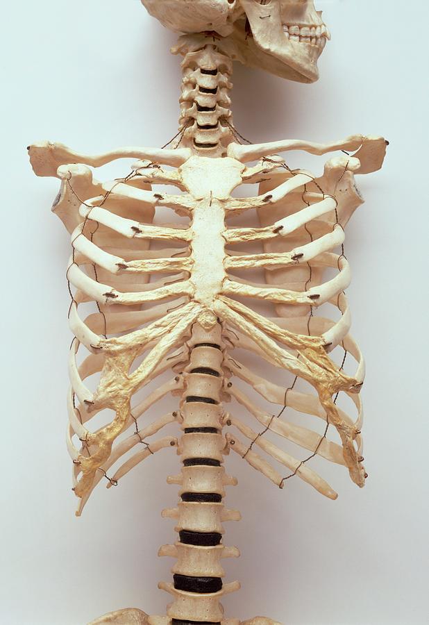
Rib Cage Of Human Body : Thoracic Cage Anatomy Body Human ... from images.fineartamerica.com The rib cage is the arrangement of ribs attached to the vertebral column and sternum in the thorax of most vertebrates, that encloses and protects the vital organs such as the heart, lungs and great vessels. In the anatomical position, the angles align with the medial border of the scapula. Together these muscles form a column, known as the erector spinae these muscles run up and down over the lower ribs and thorax (the rib cage), and cross to the low. The fibers attach to the rib cage and the pubis of the hip bones. The ribcage is made to be flexible and springy so the lungs can fill and deflate easily. Structure of a typical rib: You can click the image to magnify if you cannot see clearly. Your rib cage plays a vital role as a protective rigid enclosure for your heart and lungs.
Skeletal muscles attached to the rib cage:
Another important feature of the rib cage is the manubriosternal joint also known as the sternal angle of louis. Muscles are often named for their primary action. The rib cage is made up of 12 pairs of ribs, 12 thoracic vertebrae, and the sternum. The thoracic cage protects the heart and lungs. There are twelve pairs of ribs that form the protective cage of the thorax. Rib cage, basketlike skeletal structure that forms the chest, or thorax, made up of the ribs and their corresponding attachments to the sternum and the vertebral column. The rib cage surrounds the lungs and the heart, serving as an important means of bony protection for these vital organs. In the back, latissimus dorsi and erector spinae muscles (anatomy lesson #10) cover the 11th and 12th ribs of the thoracic cage and deeper yet are the paired abdominal kidneys flanking the. Personally, i like doing flys either flat on my back, or on an incline. The ribs are curved, flat bones which form the majority of the thoracic cage. With the upper ribs, closer to the nodule (and in the case of lower ribs, a little further from the nodule) they are curved and have a rough surface that connects them with muscles, angulus costae. Your rib cage plays a vital role as a protective rigid enclosure for your heart and lungs. The rib cage is a primarily protective structure, encircling the heart and lungs.
For more anatomy content please follow us and we think this is the most useful anatomy picture that you need. Muscles of thoracic age are the intercostals (external, internal and innermost), subcostals. Muscle spasms located in the rib cage are often observed in people who strain or overwork their upper body muscles. Anatomical illustration, images of the human body, pepin press. They are each attached to the ribs.
ribcage_by_ticor-d74f34f.jpg (1739×933) | Human rib cage ... from orig09.deviantart.net For more anatomy content please follow us and we think this is the most useful anatomy picture that you need. The costotransverse ligaments in human: It is composed of 12 pairs of ribs with their costal cartilages instead, the ribs and their small costal cartilages terminate within the muscles of the lateral abdominal wall. The rib cage surrounds the lungs and the heart, serving as an important means of bony protection for these vital organs. The tibialis anterior muscle is the largest muscle located in the anterior (front) compartment of the leg. This image added by admin. Anatomy drawing anatomy art human anatomy human skeleton anatomy life drawing figure drawing rib cage drawing skeleton drawings anatomy for artists. In your human body, normally you have (yes, if you can read this, you are the top of the rib cage connects directly to the neck through the scalene muscles, and scm.
Some of the most common causes.
Anatomical illustration, images of the human body, pepin press. It provides a strong framework onto which the muscles of the cramps in ribcage are often observed in those who strain or overwork their upper body. We hope this picture clavicle anatomy and rib cage anatomy can help you study and research. During normal breathing, contraction of the major inspiratory muscle, the diaphragm, produces both rib cage expansion and a downward movement of the diaphragm. Of or related to the morbid anatomy blog. The ribs form the main structure of the thoracic cage protecting the thoracic organs, however their main function is to aid respiration. The fibers attach to the rib cage and the pubis of the hip bones. Structure of a typical rib: There are different types of muscle, and some are controlled automatically by the the clavicular head arises from the collar bone (clavicle), while the sternocostal head arises from the breastbone (sternum) and rib cage. They are more involved in forced expiration and coughing to forcibly shrink the chest and. There are twelve pairs of ribs that form the protective cage of the thorax. How do you build muscles on your rib cage? Muscles are groups of cells in the body that have the ability to contract and relax.
The tibialis anterior muscle is the largest muscle located in the anterior (front) compartment of the leg. Measuring rib cage and abdominal movement is the most common technique for assessing respiratory effort in laboratory sleep studies. Various skeletal muscles are attached to the rib cage. Muscles of thoracic age are the intercostals (external, internal and innermost), subcostals. Together these muscles form a column, known as the erector spinae these muscles run up and down over the lower ribs and thorax (the rib cage), and cross to the low.
Human Anatomy Rib Cage And Muscles - Music Used from lh6.googleusercontent.com Seventeen muscles attach to the scapula, and it articulates with the clavicle to form the shoulder girdle or pectoral girdle, which supports movements. Rib cage anatomy and breathing. It provides a strong framework onto which the muscles of the cramps in ribcage are often observed in those who strain or overwork their upper body. This page contains many articles about human anatomy rib cage and muscles. Check out our muscle anatomy reference charts to learn faster! Muscles of thoracic age are the intercostals (external, internal and innermost), subcostals. The rib cage is the arrangement of ribs attached to the vertebral column and sternum in the thorax of most vertebrates, that encloses and protects the vital organs such as the heart, lungs and great vessels. The rib cage is a primarily protective structure, encircling the heart and lungs.
This image added by admin.
The rib cage, shaped in a mild cone shape and more flexible than most bone sets, is made up of varying elements such as the thoracic vertebra, 12 equally paired ribs, costal cartilage, and held together anteriorly by the sternum. Learn anatomy faster and remember everything you learn. Check out our muscle anatomy reference charts to learn faster! The ribs are curved, flat bones which form the majority of the thoracic cage. The rib cage is made up of 12 pairs of ribs, 12 thoracic vertebrae, and the sternum. Anatomy dissection muscle attachments of sternum, typucal ribs, atypical ribs. Muscles of the thoracic wall contain those that fill and support the intercostal spaces, those that pass between the sternum and the ribs, and those that cross several ribs between costal attachments. The costotransverse ligaments in human: They are each attached to the ribs. Various skeletal muscles are attached to the rib cage. For example, flexor, extensor, adductor and abductor are names associated with the action of the muscle. Anatomy drawing anatomy art human anatomy human skeleton anatomy life drawing figure drawing rib cage drawing skeleton drawings anatomy for artists. A rib has a flat body.
Seventeen muscles attach to the scapula, and it articulates with the clavicle to form the shoulder girdle or pectoral girdle, which supports movements rib cage muscles. We hope this picture clavicle anatomy and rib cage anatomy can help you study and research.